News – 2011/12
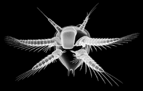
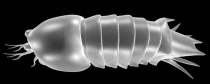
 Erugosquilla massavensis
26 December 2012:
We are happy to announce our newest paper:
Haug, C., Sallam, W. S., Maas, A., Waloszek, D., Kutschera, V. & Haug, J. T. 2012. Tagmatization in Stomatopoda – reconsidering functional units of modern-day mantis shrimps (Verunipeltata, Hoplocarida) and implications for the interpretation of fossils. Frontiers in Zoology 9, art. 31.
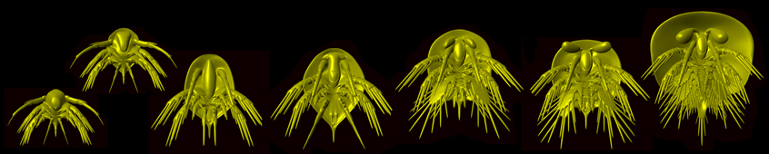
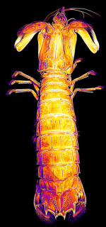
It describes morphological details of a modern mantis shrimp, Erugosquilla massavensis. Modern-day animals are important for understanding the morphology of fossils. As we are interested in fossil mantis shrimps (see, e.g., Haug et al. 2010, BMC Evolutionary Biology), a precise knowledge of their extant relatives is crucial for us.
Astonishingly, we found many details in the extant specimens that are not reported in the same way in the literature. Among these is the exact arrangement of segments of the head. The eyes and first antenna (antennula) are, according to textbooks, set off from the head shield. Yet, according to our observation not only these two segments, but also the next succeeding segment, that of the (second) antenna is set off from the remaining head. Also posterior to the head shield the segment conditions are different than expected based on literature data. The first maxilliped (leg 6) is not incorporated into the head shield, but possesses its own, but unsclerotised dorsal area. The head shield is thus only formed by the mandibular, maxillulary and maxillary segments, i.e., by three instead of five segments. Further important finds are details of the dorsal sclerites of the thorax which could explain some features of certain Jurassic mantis shrimps. Comparisons with modern larvae furthermore indicate that heterochrony, the evolutionary change of developmental timing, played an important role in the evolution towards modern mantis shrimps. The paper is Open Access and can be found here.
11 October 2012:
Our newest publication re-describing the iconic Leanchoilia superlata from the Burgess Shale is now available in its final version:
Haug, J. T., Briggs, D. E. G. & Haug, C. 2012. Morphology and function in the Cambrian Burgess Shale megacheiran arthropod Leanchoilia superlata and the application of a descriptive matrix. BMC Evolutionary Biology 12, art. 162.
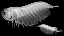
 Leanchoilia superlata
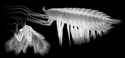
Our new findings draw a quite different conclusion about the functional morphology and thus life habits of this species than earlier reconstructions. The whole body of the animal is much more slender and flexible than previously thought. Together with relatively larger appendages than reconstructed before and a complex joint in the appendages for reducing drag forces, Leanchoilia superlata appears to have been an effective swimmer.
Furthermore, the great appendage (the first, large appendage) has more prominent spines than previously reported and is able to grasp. The second appendage is tiny compared to the others and appears to have been used especially in feeding, thus representing a kind of mouthpart. Further posterior appendages possess a massive basal part with spines on the inner side. A large membrane on the inner side should have allowed the animal to use these basal part of the appendage in a kind of “chewing motion”. Together with the presence of eyes all these new findings indicate that Leanchoilia superlata was an agile active predator, probably swimming close above the sea floor, ready for catching prey.
We also used the study of these well-preserved fossils to propose a strict scheme for describing arthropods. This scheme is thought to act as a template for future descriptions of close relatives for enhancing the comparability of such descriptions.
We have investigated about 1000 specimens of this fascinating fossil species. The specimens are housed in the Royal Ontario Museum, and were made available for study by the Curator Jean-Bernard Caron. The study was supported by the Alexander von Humboldt Foundation and the Department of Geology and Geophysics of the Yale University. The article is Open Access and can be found here.
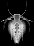
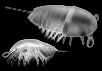
 Stadium 5 von Amphionides reynaudii
07 October 2012:
Our article on the larval development of the rare malacostracan crustacean Amphionides reynaudii was just
published:
Kutschera, V., Maas, A., Waloszek, D., Haug, C., & Haug, J. T. 2012. Re-study of larval stages of Amphionides reynaudii (Malacostraca: Eucarida) with modern imaging techniques. Journal of Crustacean Biology 32, 916–930.
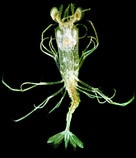
This publication does not deal with a fossil, but an extant animal. Yet, its content is so closely related to our other research, that it is presented here. Amphionides reynaudii is a very rare crustacean; until now no photographs of this species had been published. The principal approach for investigating such rare material is rather similar to the approach for reconstructing the ontogeny of fossil arthropods, another reason why this publication is presented here.
In the new publication, our co-authors and we describe different details, such as eyes and mouthparts, of various larval stages. Most images included there were recorded with composite-autofluorescence imaging. This method allows the documentation of rare museum specimens directly in their storage liquid. The investigated specimens came from the Natural History Museum in Berlin and were collected more than 100 years ago.
Among the described stages are also new, formerly unknown “stages-in-between”. One of the conclusions of the publication is therefore that the ontogeny of this crustacean is very gradual, much more gradual than previously thought. We hope that we can further amend the ontogenetic sequence in future studies, in order to compare it with the developmental sequences of other crustaceans. The paper is published here.
 Crustacean larva (nauplius) in chert
04 September 2012:
We are happy to announce the publication of our new paper on fossil crustacean larvae in chert:
Haug, C., Haug, J. T., Fayers, S. R., Trewin, N. H., Castellani, C., Waloszek, D. & Maas, A. 2012. Exceptionally preserved nauplius larvae from the Devonian Windyfield chert,
Rhynie, Aberdeenshire, Scotland. Palaeontologia Electronica 15, art. 15.2.24A.
In this paper we present all specimens (in total 79) from the collection of the Department of Geology and Petroleum Geology, University of Aberdeen, which means that each specimen is figured as a stereo image and can be viewed three-dimensionally with red-blue glasses. The crustacean larvae, in this case nauplii, were found in thin sections of chert from the Scottish Windyfield area, which is very close to the famous Rhynie chert deposits. We detected three growth stages in the material, but which only differ in size and equal each other morphologically in any other aspect. As the larvae have all only three pairs of legs and considering their small size, we suppose that these are the early stages after hatching.
The determination of the systematic position of these larvae was a bit tricky. They had previously been interpreted as anostracans (fairy shrimps), but that interpretation probably resulted from a misinterpretation of the leg morphology. After a detailed re-investigation of the legs, we suggest that the larvae might be basal branchiopods (the group including, e.g., fairy shrimps, tadpole shrimps, clam shrimps, and water fleas), but below the node of Eubranchiopoda. Another possible affiliation considering the morphology would be with Thecostraca, which includes barnacles and their relatives. In the latter case, that would be the first example of a thecostracan in freshwater, as the group is today completely marine. We hope that it will be possible to find later developmental stages of the larvae to solve the question of their exact systematic affinities. The article is Open Access and can be found here.
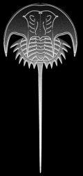 Euproops sp.
28 August 2012:
Our paper on the ontogeny of a fossil horseshoe crab was just published:
Haug, C., Van Roy, P., Leipner, A., Funch, P., Rudkin, D. M., Schöllmann, L. & Haug, J. T. 2012. A holomorph approach to xiphosuran evolution—a case study
on the ontogeny of Euproops. Development Genes and Evolution 222, 253–268.
The material is from the Carboniferous of Germany, from the Piesberg quarry near Osnabrück. The specimens are housed in the collection of the local museum (Museum am Schölerberg) and were to a large extent provided by local private collectors. The specimens could be grouped into not less than ten successive developmental stages. The smallest specimen, though incomplete, must have measured less than two millimetres (without the long telson spine), the largest one is about four centimetres long.
Especially interesting is the morphology of the posterior shield, the thoracetron, formed by the segments of the posterior body region, the opisthosoma. In young specimens the thoracetron is surrounded by spine-like extensions; in older stages the bases of these spines become more prominent and form an entire flange around the opisthosoma. What makes this ontogenetic change so interesting is the fact that the morphology seen in young specimens of the species from the Piesberg is thought to be diagnostic for a Carboniferous species of Euproops from the USA, Euproops danae. The morphology seen in older stages of the German species is thought to be diagnostic for a Carboniferous species of Euproops from the UK, Euproops rotundatus. The specimens from the Piesberg can thus not easily be distinguished from the contemporary species from the UK and USA. Only if the developmental pattern is taken into account, it is possible to properly differentiate the German morphotype from the others. From this starting point, the taxonomy of the Euproops species still needs to be revised. The paper is published here.
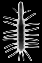 Carbotubulus waloszeki
12 August 2012:
After a long silence due to our move back to Germany, we are happy to announce now that our article on the youngest long-legged lobopodian was just published online:
Haug, J. T., Mayer, G., Haug, C. & Briggs, D. E. G. 2012. A Carboniferous non-onychophoran lobopodian reveals long-term survival of a Cambrian morphotype. Current Biology.
DOI: 10.1016/j.cub.2012.06.066
We have to admit that it does not deal with Palaeo-Evo-Devo, but still we describe a quite spectacular and unexpected find in this article. Long-legged lobopodians have until now only been found in Cambrian deposits. Our new find from the Upper Carboniferous Mazon Creek lagerstätte is 200 million years younger than the previously youngest known long-legged lobopodian. With this it is an extreme example of the long-term survival of a Cambrian morphotype. Additionally, it is preserved three-dimensionally in its concretion, and the legs were so long that they stuck out of the concretion when it was formed.
We found the specimen during a research visit at the Royal Ontario Museum Toronto in the palaeontological collections there. Together with our co-authors Georg Mayer (a specialist on velvet worms/onychophorans, modern short-legged lobopodians, at the University of Leipzig) and Derek E.G. Briggs (our host during our time at Yale University) we decided to name the new species Carbotubulus waloszeki. With this name we honour our doctoral advisor Prof. Dr. Dieter Waloszek (University of Ulm) for his work on arthropod systematics. The article is available here.
 Marrella splendens
10 March 2012:
Our article on the first marrellomorph in 'Orsten'-type preservation is accepted for print:
Haug, J. T., Castellani, C., Haug, C., Waloszek, D. & Maas, A. 2012. A Marrella-like arthropod from Cambrian of Australia: A new link between "Orsten"-type and Burgess Shale assemblages. Acta Palaeontologica Polonica.
In this paper we present new insight into faunal overlap between 'Orsten' and Burgess Shale biotas. The new marrellomorph described here, Austromarrella klausmuelleri, is represented by a three-dimensionally preserved exopod (outer branch of a leg), which exhibits traces of morphological change during ontogeny: While the proximal annuli bear a pair of lamellae characteristic for marrellomorphs, the distal annuli possess stout spines in these positions. Therefore, the spines are interpreted as ontogenetic predecessors of the lamellae. The accepted version of the article is available here.
 Spinosculda ehrlichi
02 February 2012:
On 16 January 2012 we were chosen by Hona as webseite of the week or Website van de week. We would like to thank for this honour, especially as our website is still in the early stages of development!
Hona is a Belgian association for everybody interested in fossils, minerals, geology, archaeology and nature in general. The abbreviation stands for Homo et Natura, in English: human and nature. Further information here.
 Yohoia tenuis
16 January 2012:
Our article on the ontogeny of the Burgess Shale great-appendage arthropod Yohoia tenuis and the implications on chelicerate evolution is now on Early View:
Haug, J. T., Waloszek, D., Maas, A., Liu, Y. & Haug, C. early view. Functional morphology, ontogeny and evolution of mantis shrimp-like predators in the Cambrian. Palaeontology.
DOI: 10.1111/j.1475-4983.2011.01124.x
We were able to detect ontogenetic changes in the raptorial first appendage, also called great appendage, of Yohoia tenuis. Furthermore, we found out that similar morphological differences are also present between the great appendages of two species from the Chengjiang fauna, Fortiforceps foliosa and Jianfengia multisegmentalis. While the great appendage of J. multisegmentalis looks similar to that of an earlier developmental stage of Y. tenuis, the great appendage of F. foliosa looks much more like that of a later stage of Y. tenuis. This result could indicate that the two Chengjiang species are not two separate species, but different ontogenetic stages of the same species. We also present an evolutionary scenario how great appendages evolved into chelicerae of modern chelicerates. The article is on Early View here.
 Spinosculda ehrlichi
21 December 2011:
Our article on crustacean ontogenies fossilised in limestones is finally printed:
Haug, J. T., Haug, C., Waloszek, D. & Schweigert, G. 2011. The importance of lithographic limestones for revealing ontogenies in fossil crustaceans. Swiss Journal of Geosciences 104, Supplement 1, S85–S98.
It is a summary of all our research to date on crustacean development found in lithographic limestones, amended by some new finds and an outlook on future research. As the paper had been published online already in October 2010, some of our more recent papers are not included (see Publications), for example our publication on Mesozoic mantis shrimps in 2010 in BMC Evolutionary Biology (which can be found here). The new article is published here.
 Sarotrocercus oblitus
23 November 2011:
Our new article on the Burgess Shale arthropod Sarotrocercus oblitus is finally published:
Haug, J. T., Maas, A., Haug, C. & Waloszek, D. 2011. Sarotrocercus oblitus – small arthropod with great impact on the understanding of arthropod evolution? Bulletin of Geosciences 86, 725–736.
There have been lots of different interpretations about the possible systematic position of this species and its bearing on arthropod evolution, as it seemed to have a rather strange morphology. However, it turned out to be much more "normal" than expected, as we could apply new imaging techniques not yet available for the original description. Additionally, we found two ontogenetic stages in the material, which mainly differ in size and number of segments. The article is Open Access and can be found here.
21 November 2011:
Joachim has been awarded the 13th R.J.H. Hintelmann Scientific Award of the "Freunde der Zoologischen Staatssammlung München e.V.". The ceremony will take place at the Zoologische Staatssammlung München on January 13, 2012. More information here.